Ultrasound-pros and cons
Dr Steve Roberts
What are the advantages of ULTRASOUND?
- No ionising radiation
- Portability, facilitated by laptop sized ultrasound machines.
- Simplicity, although initially US may be intimidating the common blocks e.g. femoral are readily mastered by most anaesthetists.
- Using US the nerve itself can be imaged
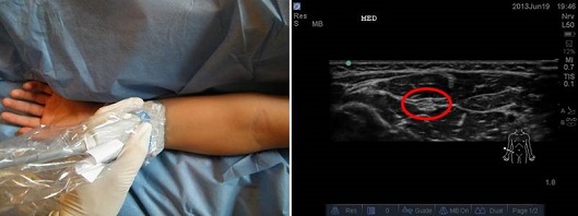
Linear probe placed in middle of forearm in transverse plane, median nerve circled in red.
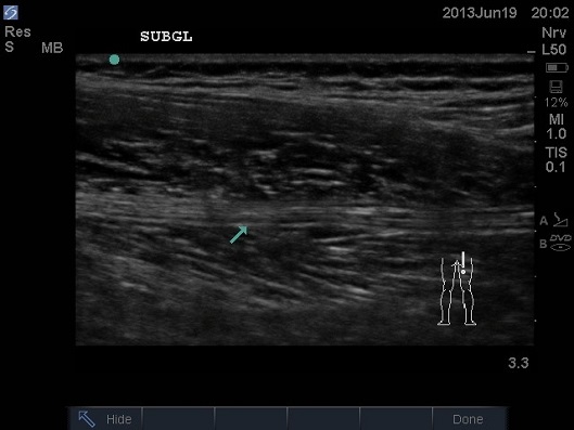
In this image the linear probe is placed in a longitudinal plane over the upper part of the posterior aspect of the thigh. The nerve is seen as a band in the middle of the screen, marked by the blue arrow.
- Not only nerves can be visualised but important fascial planes can be identified e.g rectus sheath block
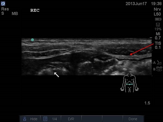
In this image the linear probe is placed in the transverse plane just inferior to the umbilicus. The white arrow indicates bowel, the red arrow indicates the correct plane in which the local anaesthetic is able to peel the muscle off the posterior rectus sheath.
- Complications from surrounding structures can be avoided, in neonates in particular vulnerable structures are very close to the areas we wish to insert our needle.
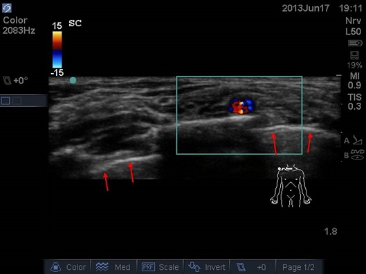
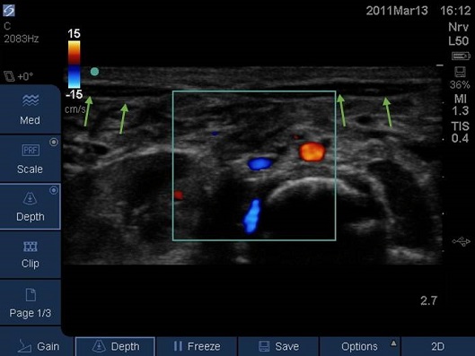
Linear probe positioned parallel and posterior to the clavicle, note the subclavian artery highlighted by Doppler anterior to the supraclavicular brachial plexus, and the pleura indicated by the red arrows.
Linear probe placed transverse over the abdomen of a neonate just caudad of the umbilicus. The aorta and inferior venae cava are highlighted by Doppler. The rectus muscle itself is only 2mm thick (green arrows), deep to it lie the abdominal contents.
- By performing a mapping or scout scan the operator creates a 3-D image of the anatomy so best needle approach can be selected.
- Real time guidance allows fine needle control and the ability to observe the local anaesthetic injectate actually spreading.
- There is evidence for faster onset and greater duration of blockade.
- Blocks can reliably be performed in the presence of neuromuscular conditions or neuromuscular blockade.
- Peripheral nerve catheter tip position can be assessed directly or indirectly by observing the injection of saline.
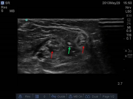
Linear probe placed on posterior aspect of lower thigh, common peroneal and tibial nerves indicated by red arrows, catheter tip indicated by green arrow.
- Most paediatric blocks are performed under general anaesthesia – this has the potential to mask warning symptoms and signs of LA toxicity and intraneural injection.
- Generally children are smaller so their nerves are very superficial; this allows higher frequency probes to be used.
- Ossification is complete by approximately 21 years of age. At birth the ossification centres of the spine are at an early stage of development; it is therefore possible to obtain excellent spinal images in neonates. With age the US windows to the spine diminish.
- The anatomy of the very young is poorly described as are the techniques associated.
- Congenital abnormalities make landmark techniques difficult to perform. And flexion deformities can prevent peripheral nerve stimulation being effective.
- Infants are particularly vulnerable to local anaesthetic toxicity, ultrasound allows doses 30-50% lower to be successfully used.
What are the disadvantages of US?
- Increased depth means a lower frequency is required for optimal imaging. As a consequence there is a lower resolution. Over time US machines have become more sophisticated, some of these machines use the returning second degree harmonic of the original frequency to produce an improved image.
- Anisotropy. Simply this means a structure is highly reflective to ultrasound. This occurs with nerves, tendons and needles. The US beam must be at or close to perpendicular to the structure for the beam to ‘bounce’ back to the probe for an image to be created. Manufacturers now have a specific program to enhance the needle image.
- Bone blocks US waves. As such imaging of the spine is increasingly difficult with increasing age.
- Artefacts are common. If a structure can only be seen in one plane it is likely to be an artefact. Manufacturers have tried to make life easier with multi-beam technology. By repeatedly changing the angle of the US beam the US filters out a lot of artefacts.
- Training. Ultrasound techniques require improved anatomical knowledge and a formal educational program.